File:Diatoms.png

Size of this preview: 734 × 600 pixels. Other resolutions: 294 × 240 pixels | 588 × 480 pixels | 940 × 768 pixels | 1,253 × 1,024 pixels | 1,400 × 1,144 pixels.
Original file (1,400 × 1,144 pixels, file size: 951 KB, MIME type: image/png)
| This free media file is from Wikimedia Commons. Its description page is included below. |
| Description |
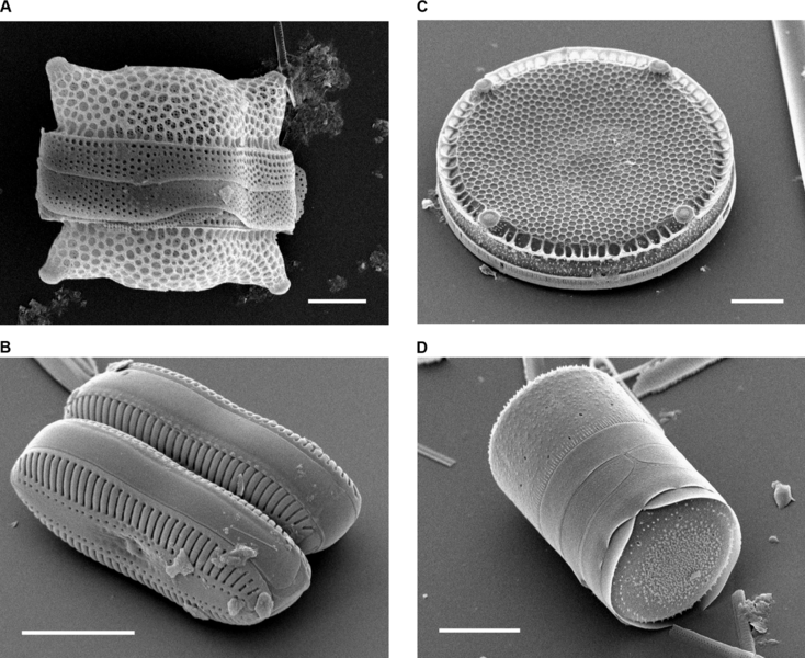
Scanning Electron Micrographs of Diatoms. (A) Biddulphia reticulata. The whole shell or frustule of a centric diatom showing valves and girdle bands (size bar = 10 micrometres). (B) Diploneis sp. This picture shows two whole pennate diatom frustules in which raphes or slits, valves, and girdle bands can be seen (size bar = 10 micrometres). (C) Eupodiscus radiatus. View of a single valve of a centric diatom (size bar = 20 micrometres) (D) Melosira varians. The frustule of a centric diatom, showing both valves and some girdle bands (size bar = 10 micrometres). |
||
| Date | Published: October 12, 2004 | ||
| Source | Bradbury J: Nature's Nanotechnologists: Unveiling the Secrets of Diatoms. PLoS Biol 2/10/2004: e306. doi:10.1371/journal.pbio.0020306 | ||
| Author | Images courtesy of Mary Ann Tiffany, San Diego State University. | ||
| Permission (Reusing this file) |
|
File history
Click on a date/time to view the file as it appeared at that time.
| Date/Time | Thumbnail | Dimensions | User | Comment | |
|---|---|---|---|---|---|
| current | 18:10, 16 November 2006 | | 1,400 × 1,144 (951 KB) | Ayacop | {{Information |Description='''Scanning Electron Micrographs of Diatoms.''' (A) ''Biddulphia reticulata''. The whole shell or frustule of a centric diatom showing valves and girdle bands (size bar = 10 micrometres). (B) ''Diploneis sp.'' This picture shows |
File usage
The following 2 pages use this file:
Global file usage
The following other wikis use this file:
- Usage on ar.wikipedia.org
- Usage on ast.wikipedia.org
- Usage on bs.wikipedia.org
- Usage on ca.wikipedia.org
- Usage on cs.wikipedia.org
- Usage on de.wikipedia.org
- Usage on en.wikipedia.org
- Usage on es.wikipedia.org
- Usage on fr.wikipedia.org
- Usage on fr.wiktionary.org
- Usage on gl.wikipedia.org
- Usage on he.wikipedia.org
- Usage on ja.wikipedia.org
- Usage on km.wikipedia.org
- Usage on nn.wikipedia.org
- Usage on pl.wikipedia.org
- Usage on pt.wikipedia.org
- Usage on sk.wikipedia.org
- Usage on test2.wikipedia.org
- Usage on zh.wikipedia.org